
Synergetic Neuro-Fuzzy Feature Selection and Classification of Brain Tumors
- 0 Collaborators
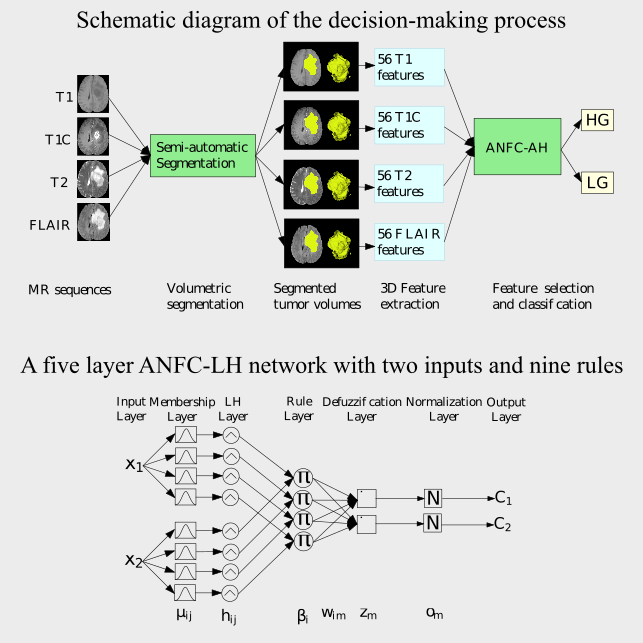
Brain tumors constitute one of the deadliest forms of cancers, with a high mortality rate. Of these, Glioblastoma multiforme (GBM) remains the most common and lethal primary brain tumor in adults. Tumor biopsy being challenging for brain tumor patients, noninvasive techniques like imaging play an important role in the process of brain cancer detection, diagnosis and prognosis; particularly using Magnetic Resonance Imaging (MRI). Therefore, development of advanced extraction and selection strategies of quantitative MRI features become necessary for noninvasively predicting and grading the tumors. In this paper we extract 56 three-dimensional quantitative MRI features, related to tumor image intensities, shape and texture, from 254 brain tumor patients. An adaptive neuro-fuzzy classifier based on linguistic hedges (ANFC-LH) is developed to simultaneously select significant features and predict the tumor grade. ANFCLH achieves a significantly higher testing accuracy (85:83%) as compared to existing standard classifiers. Features selected by the system also perform better than those chosen using well-known feature selection strategies. ...learn more
Project status: Published/In Market
Intel Technologies
Other
Overview / Usage
Improving lifespan has resulted in an ageing population, with several diseases including cancer becoming a leading cause of death worldwide. Glioblastoma multiforme (GBM) is the most common and lethal primary brain tumor in adults. Noninvasive techniques like imaging play an important role in the process of brain cancer detection, diagnosis and prognosis. Innovation and improvement of medical imaging equipments, over the past decade, have enabled us to move towards quantitative imaging based treatment planning; particularly using Magnetic Resonance Imaging (MRI).
MRI is routinely used in the diagnosis, characterization, and clinical management of gliomas. It is able to capture multidimensional in vivo portraits of GBMs by simultaneously extracting structural, compositional, and physiological information. It is also safe, as it does not involve any exposure to radiation. While repeated tumor biopsies are challenging in brain tumor patients, MRI has the added advantage of noninvasively capturing the heterogeneity of gliomas Feature extraction from the ROI/VOI has been reported in literature, encompassing tumors of different organs. Texture and shape features, involving gray-level co-occurrence, run-length and morphological features, were extracted from lung CT images for differentiating between benign and malignant mediastinal nodes in lung cancer. Computer-aided systems for detection/diagnosis of breast cancer, based on imaging features extracted from mammography and ultrasound images, are also available. 2-dimensional quantitative MRI imaging features have been studied for tumor classification. The objective of our research is to extract an optimal set of quantitative 3 dimensional MRI features from the tumor volume of interest (VOI), and use them to effectively discriminate between the categories HG and LG.
Since the features extracted from MR images involve certain unknown statistical properties, and the class boundaries are not always well-defined, therefore standard classifiers do not perform satisfactorily. Although fuzzy rule based classifiers demonstrate good performance in this scenario, the proper selection of its parameters often requires maneuvering. Neural networks were used to automatically learn the parameters in a neuro-fuzzy framework. A neural network constructed from fuzzy classification rules, with parameters adaptively learned through training, is called an adaptive neuro-fuzzy classifier (ANFC). It thereby combines the merits of both fuzzy sets and neural networks.
As classification accuracy may be affected by overlapping classes in the input feature space, one way to enhance performance is by projecting the features to a different domain. However this leads to loss of semantics of these features. A similar result can be achieved by controlling the contribution of each feature to the classification process. Those features that cause the overlap can be reduced using the concept of linguistic hedge (LH).
Methodology / Approach
In this paper we propose an automated decision-making system capable of extracting an optimal set of 3-dimensional quantitative MRI features from the patient MR image sequences. It can effectively discriminate between the brain tumor categories (HG or LG). We use an adaptive neuro-fuzzy classifier with linguistic hedges (ANFC-LH), for simultaneous feature selection and classification. The system employs quantitative imaging features based on gray level statistics, texture, run length, and shape, extracted from the VOI (as marked by medical experts) from 3-dimensional MR images.
Technologies Used
The model was developed using python 2.7 with scikit-learn on a with an Intel Core i7 - 2600 K, 3.40 GHz processor having 16 GB RAM.